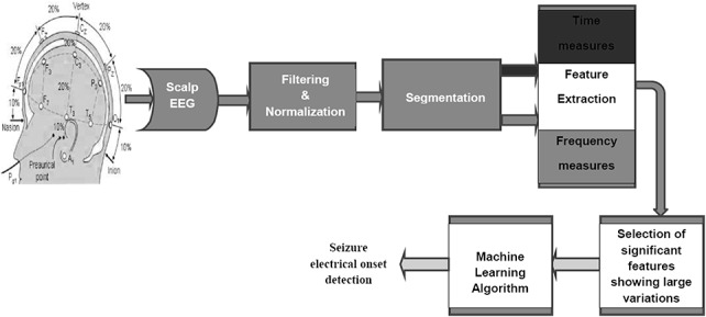
FIG. 2.

Steps in EEG data collection and processing for the automated seizure detection system. The image of the 10 to 20 electrode placement system used in this figure is adapted from Bioelectromagnetism, Principles and Applications of Bioelectric and Biomagnetic Fields (page no. 368) by J. Malmivuo and R. Plonsey, 1995, Oxford University Press, New York, adapted with permission.52
