Figure 7.
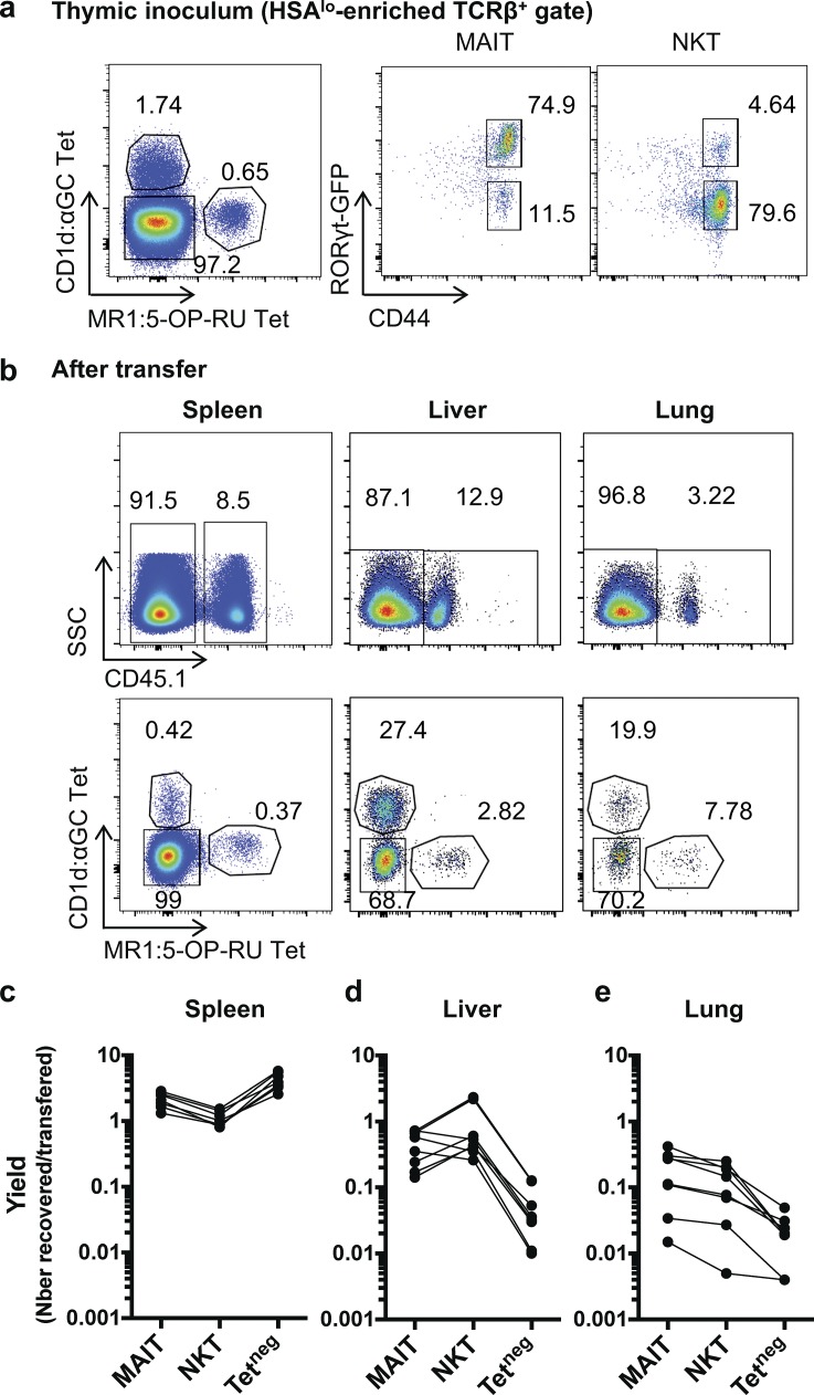
NKT and MAIT cells from the thymus are more prone to locate to the liver and to the lungs than conventional T cells. Mature enriched (HSAlow) CD45.1+ thymocytes were transferred into CD45.2+ hosts, and the indicated organs were analyzed 36 h later for NKT (CD1d:PBS57Tet+), MAIT (MR1:5-OP-RU Tet+), and conventional T cell numbers. (a) example of staining of the inoculum (HSAhi-depleted thymocytes). (b) Examples of staining in the indicated organs after transfer. (c–e) Yield (no. recovered/no. injected cells) of the indicated subsets in the spleen (c), the liver (d), and the lungs (e; n = 6 transferred mice; three independent experiments).

