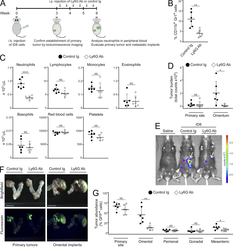
Figure 3.
Neutrophil depletion decreases omental metastasis. Immunocompetent C57BL/6 mice were injected i.b. with ID8 cells that express GFP and luciferase. At 3 wk thereafter, formation of primary tumors was confirmed by bioluminescence imaging. Mice were then randomized into groups that were administered either Ly6G Ab or control Ig twice a week for 3 wk (n = 6 mice per group). Following Ab treatment, peripheral blood and tumors in each mouse of each group were analyzed. (A) Experimental scheme. (B) Abundance of neutrophils in peripheral blood, quantified by flow cytometric analysis. **, P < 0.01 (unpaired t test). (C) Analysis of peripheral blood by CBC test. ****, P < 0.0001 (unpaired t test). (D) Sizes of primary tumors and omental implants detected by bioluminescence imaging and calculated from emitted signals. *, P < 0.05 (unpaired t test). (E) Representative images of mice with primary tumors (arrowheads) and omental implants (arrows) detected by bioluminescence imaging. (F) Representative images of tissues viewed under light and fluorescence microscopy. Bars, 10 mm. (G) Abundance of tumor in tissues of the primary site (left ovary and oviduct) and in visceral fat tissues, quantified by flow cytometric analysis of GFP+ cells. *, P < 0.05; **, P < 0.01 (unpaired t test). Error bars in B–D and G represent SD.

