Figure 7.
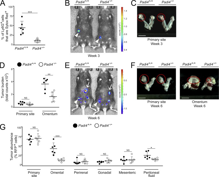
Omental metastasis is decreased in NET-defective, neutrophil-specific, Padi4-deficient mice. Female neutrophil-specific Padi4−/− mice and control siblings were injected i.b. with ID8 cells that express RFP and luciferase and thereafter were evaluated for NET formation and tumor progression. (A) At 3 wk, mice in both groups were euthanized (n = 6 mice per group). Fresh omental tissues were stained with Ly6G Ab and Sytox Red dye, and staining was analyzed by flow cytometry. Shown is the proportion of omental Ly6G+ cells that were Sytox Red+ in each mouse of each group. ***, P < 0.001 (unpaired t test). (B and C) Representative examples of primary tumors detected at 3 wk by bioluminescence imaging (arrowheads; B) and by light microscopy (circled; C). Bar, 10 mm. (D–G) Analysis of tumor progression at 6 wk (n = 6 mice per group). (D) Sizes of primary tumors and omental implants detected in each mouse by bioluminescence imaging. **, P < 0.01 (unpaired t test). (E) Representative images of mice with primary tumors (arrowheads) and omental implants (arrows). (F) Representative images of tumors viewed under light microscopy (circled). Bars, 10 mm. (G) Abundance of RFP+ tumor cells in tissues of the primary site, visceral fat tissues, and peritoneal fluid in each mouse, quantified by flow cytometric analysis. *, P < 0.05; ***, P < 0.001 (unpaired t test). Error bars in A, D, and G represent SD.

