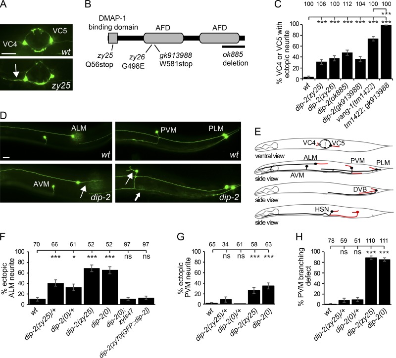
Figure 1.
dip-2 regulates neuronal morphology and migration. (A) VC4 and VC5 neurons in WT and a zy25 mutant. Arrow points to ectopic neurite from VC4. (B) DIP-2 protein schematic showing domain organization and mutations. (C) Quantification of VC morphology defects in Dip-2 mutants. (D) Mechanosensory neuron images. (E) Worm schematics summarizing dip-2 neuronal morphology and migration defects (red). (F–H) Quantification of Dip-2 neuronal morphology defects in ALM (F) and PVM (G and H). dip-2 mutants display ectopic neurites (arrows) from cell bodies and axon branching (thick arrow in D) defects. Bars, 20 µm. Error bars indicate SEM of proportion (n = 51–112). Significance compared with WT using one-way ANOVA with Tukey post hoc test. *, P < 0.05; ***, P < 0.001.

