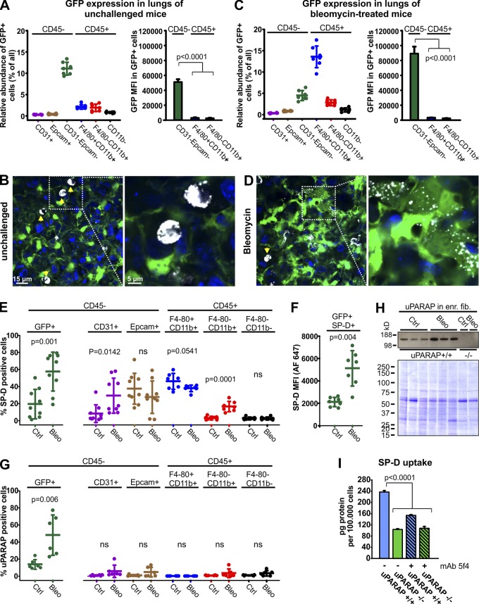
Figure 4.
Uptake of SP-D in uPARAP-positive fibroblasts in vivo after lung injury. (A and C) Flow cytometry–based quantification of GFP-positive cells and their MFIs in lung cell populations acquired from unchallenged (A) or bleomycin-treated (C) col1a1.GFP mice. (B and D) Uptake of Alexa Fluor 647–labeled SP-D (white) by lung cells was examined using confocal imaging 24 h after intratracheal instillation into the lungs of unchallenged or bleomycin-treated col1a1-GFP mice. SP-D uptake in unchallenged mice (B) was dominated by alveolar macrophages (arrows), but after bleomycin injury (D), fibroblasts (green) with SP-D uptake were abundant. Hoechst 33342 was used for visualization of cell nuclei (blue). (E–G) Flow cytometry–based analysis of ligand uptake by lung cells from unchallenged or bleomycin-treated col1a1-GFP mice. Cellular uptake of fluorescent ligands was recorded 24 h after intratracheal instillation of Alexa Fluor 647–labeled SP-D (E and F), or 24 h after i.v. injection of Alexa Fluor 647–labeled anti-uPARAP mAb2h9 (G). (E) Fraction of GFP+ fibroblasts (left) and other lung cell populations (right) positive for SP-D uptake. Col1a1-GFP mice that had not been injected with fluorescent SP-D were used as negative controls. (F) Level of SP-D associated with SP-D+GFP+ fibroblasts as determined by Alexa Fluor 647 MFI. (G) Fraction of GFP+ fibroblasts (left) and other lung cell populations (right) positive for uptake of fluorescent mAb 2h9. Col1a1-GFP mice injected with Alexa Fluor 647–labeled irrelevant murine IgG was used as negative control. n = 7–9. Two-tailed Student’s t test was used to test significance. (H) Top: Western blotting for uPARAP in lysates prepared from untreated or bleomycin-treated mouse lung cells enriched for fibroblasts (see Materials and methods). Cell lysates from uPARAP-deficient mice (−/−) were included as negative controls. Bottom: Nonreducing SDS-PAGE and Coomassie Brilliant Blue staining of protein lysates as loading controls for the Western blot. (I) Internalization of radiolabeled SP-D in primary dermal fibroblasts from uPARAP−/− mice and WT (uPARAP+/+) littermates. Analysis was performed as in Fig. 3 F.

