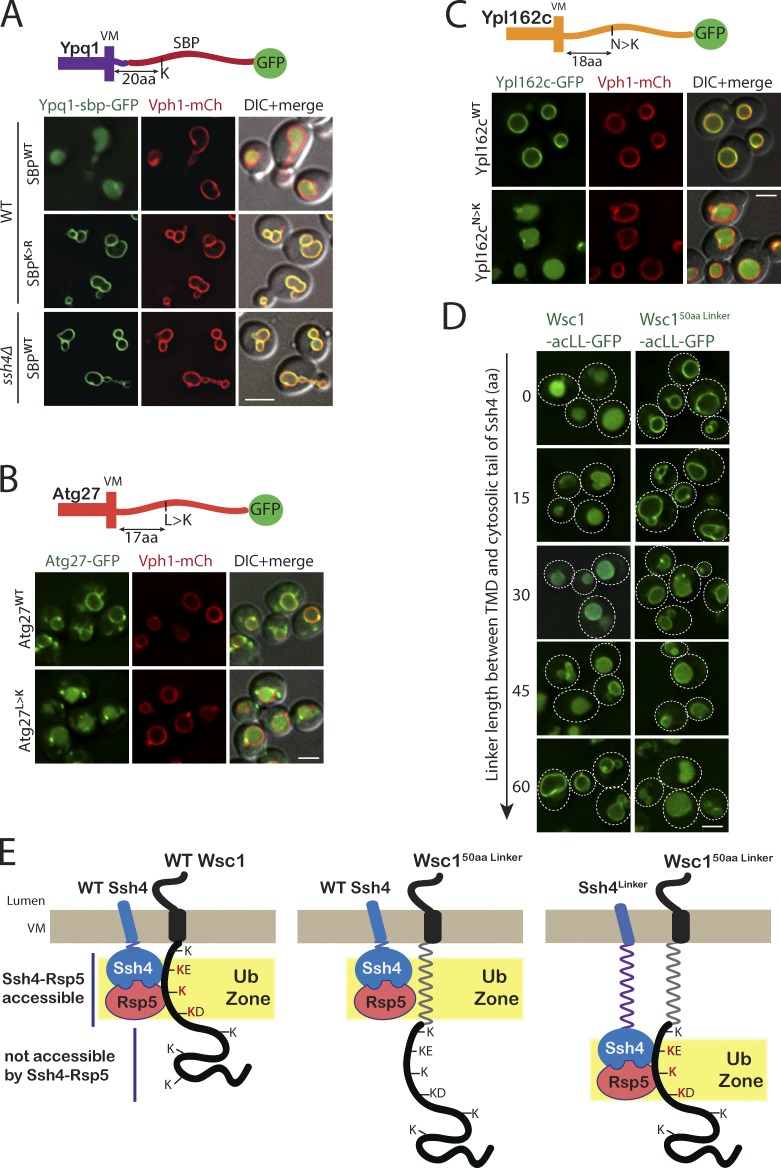
Figure 7.
Addition of a lysine to the ubiquitination zone results in constitutive degradation of stable VM proteins. (A) Fluorescence microscopy of WT and ssh4Δ mutant cells expressing Ypq1-SBPWT or SBPK>R-GFP. Scale bar: 2 µm. VM marker, Vph1-mCherry. (B) WT cells expressing GFP tagged Atg27WT or Atg27L>K mutant. (C) WT cells expressing GFP tagged Ypl162cWT or Ypl162cN>K. (D) WT cells expressing Wsc1-acLL-GFP or Wsc150aaLinker-acLL-GFP and Ssh4 with linkers of varying lengths (15, 30, 45, and 60 aa) between the TMD and the cytosolic domain. Dashed lines indicate the cell periphery. Scale bar: 2.5 µm. VM marker, Vph1-mCherry. (E) Model for Ssh4–Rsp5–mediated recognition of cytosolic lysines in the ubiquitination zone in cargos at the VM.

