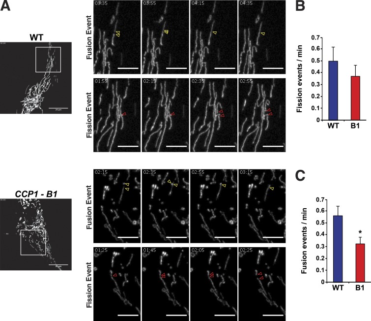
Figure 4.
Retinal pigmented epithelial cells lacking CCP1 undergo reduced mitochondrial fusion. (A) Representative examples of mitochondrial fission and fusion events in WT RPE1 cells and in a CCP1 null (CCP1-B1) RPE1 cell line. Red arrowheads indicate a fission event, while yellow arrowheads highlight a fusion event. Left (lower power): bars, 20 µm. All other panels present higher magnification of respective insets followed at 20-s intervals; bars, 5 µm. (B) We counted mitochondrial fission events from live cell imaging in A and calculated the fission rate as fission events per minute for WT RPE1 cells and in a CCP1 null (B1) RPE1 cell line. (C) We counted mitochondrial fusion events from live-cell imaging in A and calculated the fusion rate as fusion events per minute for WT RPE1 cells and in a CCP1 null (B1) RPE1 cell line. t test: *, P < 0.05. n ≥ 10 cells per genotype, and n = 3 independent experiments. Error bars are SEM.

