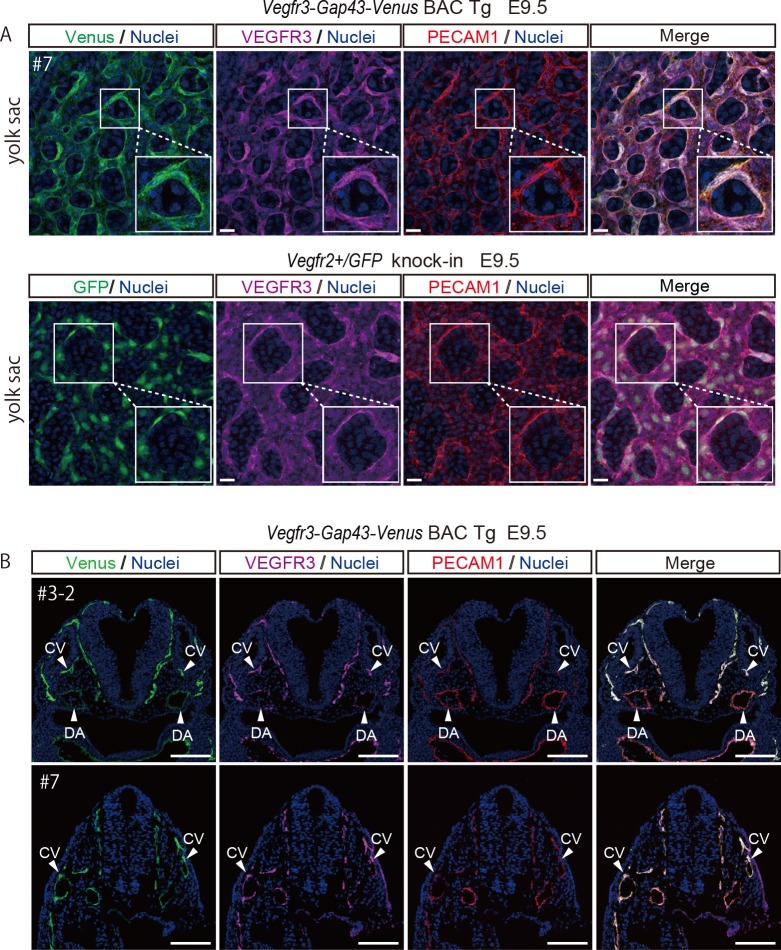
Fig 3. Membrane localization of Venus in a Vegfr3-Gap43-Venus BAC Tg yolk sac at E9.5 and Venus expression in a Vegfr3-Gap43-Venus BAC Tg embryo at E9.5.
(A) Immunofluorescence analysis of the Vegfr3-Gap43-Venus BAC Tg yolk sac at E9.5 for a vascular endothelial marker, PECAM1 (red), Venus (anti-GFP, green), VEGFR3 (magenta) and Nuclei (Hoechst33342, blue). Note that the Venus of the Vegfr3-Gap43-Venus BAC Tg yolk sac was localized in the plasma membrane of endothelial cells, while the GFP of Vegfr2+/GFP knock-in was localized in the cytoplasm. Scale bar: 20 μm. All images were captured by a Leica TCS-SP8 confocal microscope using a 40x/1.25 oil objective lens. (B) Immunofluorescence images of the Tg embryo at E9.5 for PECAM1 (red), Venus (anti-GFP, green), VEGFR3 (magenta) and Nuclei (Hoechst33342, blue). Cryosections were prepared from the Tg embryos and subjected to immunohistochemistry. Note that endogenous VEGFR3 and Venus were overlapped in the vascular endothelial cells of the Tg embryo. DA: dorsal aorta; CV: cardinal vein. Scale bar: 100 μm. All images were captured by a Leica TCS-SP8 confocal microscope using a 20x/0.7 dry objective lens.

