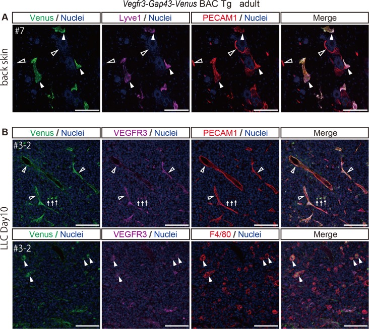
Fig 5. Venus expression in the normal back skin and tumor of Vegfr3-Gap43-Venus BAC Tg adult mice.
(A) Immunofluorescence images of the back skin of an adult Tg mouse for Venus (anti-GFP, green), Lyve1 (magenta), PECAM1 (red) and Nuclei (Hoechst33342, blue). Closed and open arrowheads indicate Lyve1-positive lymphatic endothelial cells and Lyve1-negative vascular endothelial cells, respectively. (B) Immunofluorescence images of the tumor implanted in the Tg for Venus (anti-GFP, green), VEGFR3 (magenta), PECAM1 (upper panel) or F4/80 (lower panel) (red) and Nuclei (Hoechst33342, blue). Open arrowheads indicate PECAM1-positive vascular endothelial cells. Arrows indicate PECAM1-negative cells with a round morphology. Closed arrowheads indicate F4/80-positive macrophages. Scale bar: 100 μm. All images were captured by a Leica TCS-SP8 confocal microscope using a 20x/0.7 dry objective lens.

