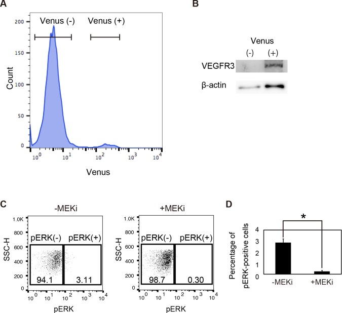
Fig 8. Detection of pERK signaling of vascular endothelial cells in Vegfr3-Gap43-Venus Tg embryos at E9.5.
(A) Flow-cytometry analysis of Vegfr3-Gap43-Venus Tg embryos at E9.5. (B) Western blot analysis of Venus-positive and -negative cells from Tg embryos. (C) Phosphorylated ERK signaling in Venus-positive cells cultured in the presence or absence of the MEK inhibitor. (D) Percentage of pERK-positive cells in Venus-positive cells.

