
Figure 1.
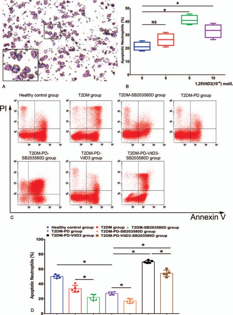
Morphology of isolated peripheral blood neutrophils; 1,25VitD3-induced apoptosis of peripheral blood neutrophils from patients with T2DM and PD in a dose-dependent manner, and flow cytometry of 1,25VitD3-induced apoptosis of neutrophils in each group. Peripheral blood neutrophils were stained with Wright's stain and presented a slightly red color in which many particles distributed in the cells presented a neutral pink and purple color. The nuclei of neutrophils exhibited a multilobulated shape. Large amounts of neutral nonbasophilic and noneosinophilic granules were observed in the cytoplasm of neutrophils. The nuclei had a characteristic lobed appearance, mostly containing a nucleus divided into 2 to 5 lobes; multiple nuclei fusions were commonly observed (Fig. 1A). (Original magnification 200 × , scale bar in Fig. 1A: 200 μm). Neutrophils were cultured for 24 hours in the presence of 0, 1 × 10⁻⁶, 1 × 10⁻⁸, or 1 × 10⁻¹⁰ mol/L 1,25VitD3 as indicated, and neutrophil apoptosis was assessed by flow cytometry. Treatment with 1,25VitD3 at the dose 1 × 10⁻⁸ mol/L induced the highest rate of neutrophil apoptosis compared with the other groups (Fig. 1B) (∗P <.05, NS: P>.05). A total of 1 × 10⁶ of peripheral blood neutrophils were pretreated with 10 μmol/L SB203580 in the T2DM-PD, T2DM-PD-SB203580, and T2DM-PD-VitD3-SB203580 groups for 0.5 hours, and neutrophils in the T2DM-PD-VitD3-SB203580 group were then incubated with 1 × 10⁻⁸ mol/L 1,25VitD3 for 24 hours. According to the analysis data, the RUQ represents the terminal apoptotic and necrotic neutrophils (FITC [+], PI [+]). The RLQ represents the early apoptotic neutrophils with positive Annexin V and negative PI (FITC [+], PI [-]). The LLQ represents the vital and living neutrophils with negative Annexin V and PI expression (FITC [+], PI [-]). The counted numbers of apoptotic neutrophils in the right lower quadrant are displayed (Fig. 1C). The total cell counts are shown as the % of 106 cells. Neutrophils in the T2DM-PD-VitD3 group expressed the highest apoptotic level among all of the groups, whereas neutrophils in the T2DM-PD-SB203580 group expressed the lowest apoptotic level (Fig. 1D). The data are presented as the mean ± std (∗P <.05, NS: P >.05). 1,25VitD3 = 1,25-Dihydroxyvitamin-D3, FITC = fluorescein isothiocyanate, LLQ = lower left quadrant, PD = periodontitis, PI = propidium iodide, RLQ = right lower quadrant, RUQ = right upper quadrant, T2DM = type 2 diabetes mellitus.