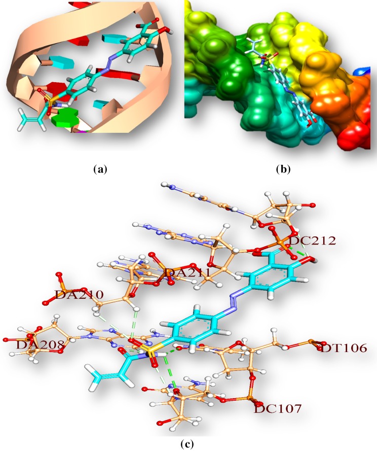
Fig. 9.
M1 interaction with Tuberculosis’s DNA in minor groove. a Drug is represented by stick and double helical DNA structure is represented by ladder and rings, b double helical structure of DNA represented by M1 represented by stick model and are coloured according to elements, c interactions of ligand with DNA base pairs (A, T, G, C); the interaction types. Hydrogen bonds are in green. Ligand surrounding base pairs are in three letters code represented in black

