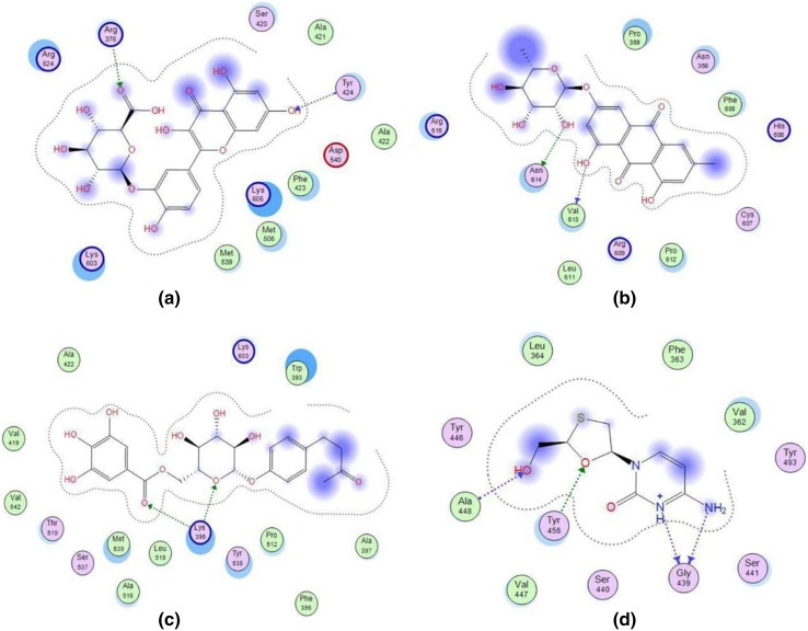
Fig. 3.
Schematic configuration of docking results of Quercetin-3-glucuronide (a), Frangulosid (b), Lindleyin acid (c) and Lamivudine (d) with binding pocket of HBV DNA polymerase. Polar residues are shown in pink. Acidic residues have been circled in red color while basic ones are encircled by blue color. Acceptance or donation of electron is shown by the respective arrow pointing towards or away from the residue. Dashed line shows the proximity contour

