
Fig. 4. Therapeutic effect of mastoparan is specific to MC activation.
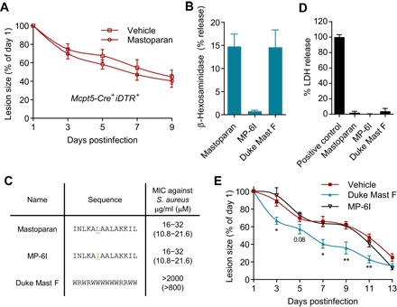
(A) Quantification of lesion sizes at indicated days after infection of MC-depleted mice and vehicle or mastoparan treatment (n = 6 to 7). Note that healing rates in MC-depleted mice were slower than that in MC-sufficient mice due to larger initial lesions. Data represent two independent experiments. (B) Degranulation of MC/9 cells expressed as β-hexosaminidase release by 25 μM mastoparan, MP-6I, and Duke Mast F. (C) Amino acid sequence and MIC (minimum inhibitory concentration) for mastoparan or each of its analogs. (D) Cytotoxicity of mastoparan or its analogs measured by LDH assay 4 hours after incubation of L929 cells at a concentration of 50 μM. (E) Quantification of lesion sizes in each treatment group (n = 5). Data were analyzed via unpaired two-tailed Student’s t test or one-way ANOVA. Error bars represent SEM. *P < 0.05, **P < 0.01.