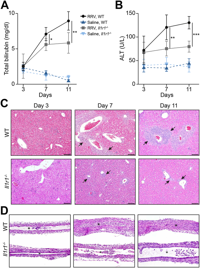
Fig. 2. Loss of IL-1R1 improves cholestasis and tissue injury.
(A and B) Serum total bilirubin and ALT levels in WT and Il1r1−/− mice. N=3–7 per group/time point; *P <0.05, **P <0.01, ***P < 0.001. Representative sections stained with H&E showing improvement in portal inflammation (liver, panel C) and restoration of duct epithelium and patent lumen (EHBD, panel D) in Il1r1−/− mice after RRV (compared to WT controls). N=13 for Il1r1−/− mice; N=15 for WT mice. Arrows denote portal tracts; *Denotes duct lumen; scale bar: 100μm; magnification=100× for EHBD.

