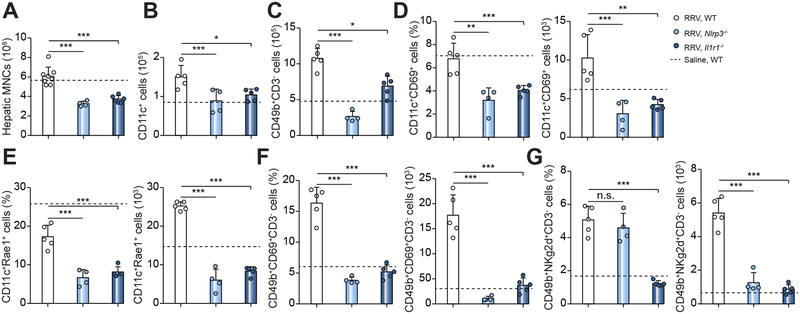
Fig. 6. Population and activation of DCs and NK cells in neonatal livers after RRV infection.
Flow cytometric analysis of immune cells from livers at 3 days showing the lower number of hepatic total mononuclear cells (A), dendritic cells (CD11c+) (B), and NK cells (CD49b+) (C). The activation of DC and NK is blunted with lower percentage of DCs positive for CD69+ (D) or RAE1+(E), and NK cells positive for CD69+ (F) or NKg2d+ (G). N=4–8 livers per group. *P < 0.05, **P < 0.01, ***P < 0.001. The dash line represents values of WT mice injected with normal saline.

