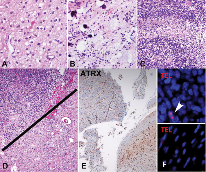
Figure 2.

Pilocytic astrocytoma with anaplasia (PA‐A) with ATRX loss and ALT in the anaplastic component only (case 1). The tumor at first resection was hypocellular, composed of piloid areas with oligodendroglial‐like cells and Rosenthal fibers. A. The same component was present in a subsequent resection, in the absence of radiotherapy B, but in addition contained sharply demarcated hypercellular areas with brisk mitotic activity and pseudopallisading necrosis C consistent with spontaneous anaplastic transformation. Distinct anaplastic areas in upper fields D, contained ATRX loss E and ultrabright foci on telomere FISH indicating ALT (arrow) F, which were absent in the lower grade PA in lower fields D‐F, as well as in the PA precursor two years prior (not shown).
