Fig. 1.

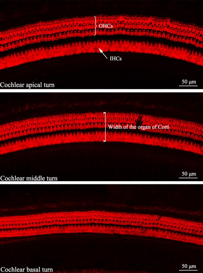
Representative images of surface preparations of the New Zealand White (NZW) rabbit cochlea (× 400, TRITC-conjugated phalloidin staining). Three rows of outer hair cells (OHCs) and the single row of inner hair cells (IHCs) are visible

Representative images of surface preparations of the New Zealand White (NZW) rabbit cochlea (× 400, TRITC-conjugated phalloidin staining). Three rows of outer hair cells (OHCs) and the single row of inner hair cells (IHCs) are visible