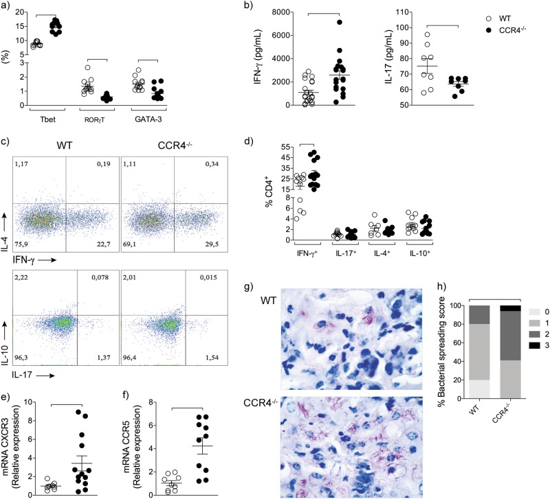
Fig. 4. Absence of CCR4 exacerbates lung Th1 inflammation and bacterial spreading into the lungs.
WT (white circles) and CCR4−/− (black circles) mice were infected with M. tuberculosis as described in the Fig. 1. At 70 days of infection, the lungs were collected. Frequency of lung CD4+Tbet+, CD4+RORγt+ and CD4+GATA-3+ cells a. IFN-γ and IL-17 levels in the lung homogenates b. Representative analysis of IFN-γ-, IL-4-, IL-17- and IL-10-producing CD4+ cells c. Frequency of IFN-γ-, IL-4-, IL-17- and IL-10-producing CD4+ cells d. CXCR3 e and CCR5 f gene expression in the lung homogenates. Representative Ziehl-Neelsen staining on lung sections at 70 days of infection (magnification, ×400) g. Lung bacterial spreading score h. Data are representative of three-independent experiments (n = 9–22) expressed as the mean ± SEM. Symbols represent individual animals and bars show the difference (P < 0.05) between the groups

