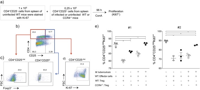
Fig. 5. CCR4 regulates the suppressor function of regulatory T cells during M. tuberculosis infection.
WT (white circles) and CCR4−/− (black circles) mice were infected with M. tuberculosis as described in the Fig. 1 or left uninfected. At 70 days of infection, the spleens were collected. CD4+CD25− (effector) cells (1 × 10⁵) purified from the spleens of uninfected WT mice were co-cultured with CD4+CD25+ (regulatory) cells (0.25 × 10⁵) from the spleens of uninfected or infected WT or CCR4−/− mice. Co-cultures were stimulated with ConA and after 96 h, proliferation was assessed a. Foxp3 expression on CD4+CD25+ cells b, c. Proliferation was assessed by the Ki-67 expression on CD4+CD25− cells d, e. As a positive control, CD4+CD25− cells were stimulated with ConA in the absence of CD4+CD25+ cells (gray circles). Data from two reproduced experiments #1 and #2 (n = 3–5) expressed as the mean ± SEM. Symbols represent individual animals and bars show the difference (P < 0.05) between the groups

