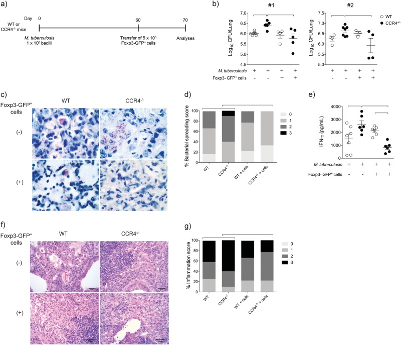
Fig. 6. Foxp3-GFP+ cell transfer renders CCR4−/− mice more resistant to M. tuberculosis infection and ameliorates pulmonary inflammation.
WT (white bars) and CCR4−/− (black bars) mice were infected with M. tuberculosis as described in the Fig. 1. Spleen Foxp3-GFP+ cells (5 × 10⁵) were transferred by the intra-tracheal route to day 60-infected WT or CCR4−/− mice. As an experimental control, infected mice were left without cell transfer. Ten days after Foxp3-GFP+ cell transfer, lungs were evaluated a. Lung CFU numbers from two reproduced experiments #1 and #2 (n = 3–7) expressed as the mean ± SEM b. Ziehl-Neelsen staining c and lung bacterial spreading score d. IFN-γ levels in the lung homogenates e. Data are representative of two-independent experiments (n = 6–7) expressed as the mean ± SEM. Representative analysis of lung inflammation f and inflammation score g of WT and CCR4−/− mice were obtained from lung sections stained with hematoxylin and eosin. Magnification ×400. Symbols represent individual animals and bars show the difference (P < 0.05) between the groups

