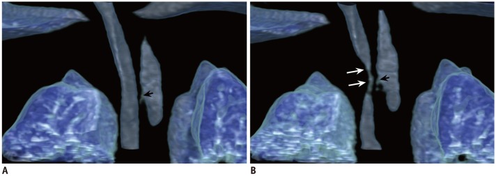
Fig. 8. Tracheomalacia in 2-month-old boy with tetralogy of Fallot after esophagoesophagostomy for esophageal atresia and ligated tracheoesophageal fistula.
Coronal inspiratory (A) and expiratory (B) volume-rendered CT images show severe expiratory tracheal collapse (long arrows) indicating tracheomalacia at thoracic inlet level. Longitudinal extent of focal tracheomalacia is nicely illustrated on expiratory-phase 4D CT image (B). Remnant of ligated tracheoesophageal fistula (short arrows) is noted.

