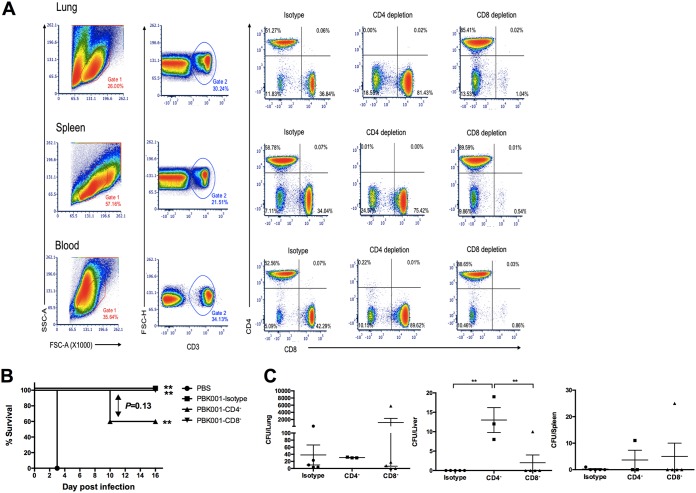
FIG 4.
Role of T cell populations in PBK001 vaccine-induced protection. (A) Confirmation of T lymphocyte depletion using three-color FC analysis of cells from intact, CD4-depleted, and CD8-depleted mice. Cells (106) from lung, spleen, or blood were stained with APC-anti-CD3, PE-anti-CD4, and PerCP Cy5.5-anti-CD8 antibodies after in vivo depletion. Results are shown on density plots with a logarithmic scale. The percentages of T lymphocytes from each organ of isotype and depleted sets are shown in each quadrant. (B) C57BL/6 mice (n = 5/group) were immunized with strain PBK001 or PBS. Two weeks after the second boost, mice were treated with rat IgG2b isotype control, anti-mouse CD4 and anti-mouse CD8α and then challenged with 3 LD50 of B. pseudomallei K96243. Mice were monitored daily and survival (percent) differences were determined using a log rank (Kaplan-Meier) test. (C) Bacterial burden in lung, liver, and spleen was determined on day 16. The difference of CFU/organ from each was compared using one-way ANOVA (Tukey’s test). Bars represent means with standard errors of the means (SEM). **, P < 0.01.

