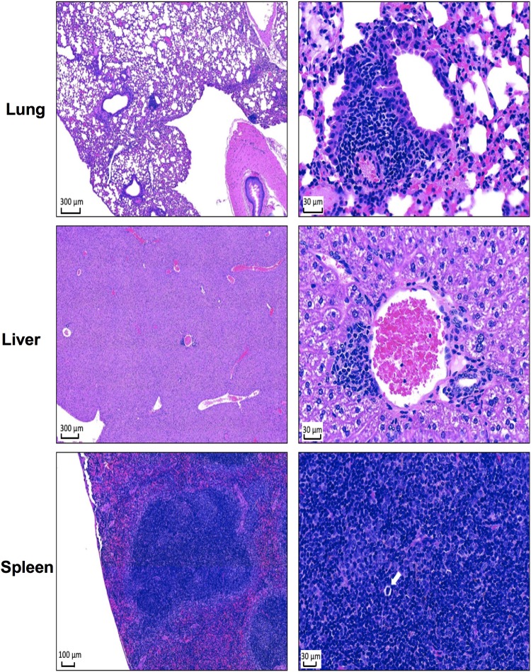
FIG 5.
Histopathology analysis of PBK001-vaccinated mouse tissues after aerosol challenged with B. pseudomallei K96243. At the end of study, organs were harvested from three mice. The tissues were fixed and stained with H&E. Stained tissues display findings seen in lung, liver, and spleen 27 days after challenge. The left figures represent images of 4× (bar = 300 μm) (lung and liver) and 10× (bar = 100 μm) (spleen) magnification, and the right figures represent 40× (bar = 30 μm) magnification. The white arrow indicates a hematopoietic lesion.

