
Figure 1.
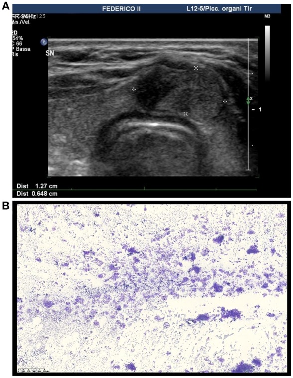
Ultrasound and cytological features of the thyroid nodule in our patient. (A) Ultrasound image showing an isoechoic solid nodule with a hypoechoic cranial component with blurred margins located in the isthmus of the thyroid. (B) Medium power magnification showing a hypercellular smear featuring thyrocytes arranged in microfollicular structures (DiffQuik staining, 100X).