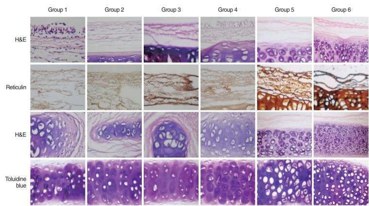
Fig. 2.
Sections of decellularized tracheas from groups 1–6. First two rows depict H&E and reticulin staining of epithelial and primary mesenchymal zones (basal membrane, collagen fibers, vesicles, and fibroblasts); third and fourth rows depict H&E and toluidine blue staining of secondary mesenchymal zone (matrix). Magnification, ×200, respectively.

