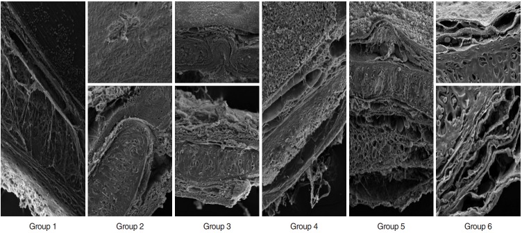
Fig. 3.
Scanning electron microscopy examination of sections from decellularized tracheas of groups 1–6. Group 1: deformed cells on luminal surface, preserved basal membrain (BM), dispersed collagen fibers; preserved matrix, deformed chondrocytes (magnification, ×500). Group 2: no cells on luminal surface; affected BM, compact collagens, deformed isogenic groups, acellular (magnification, ×350 and ×600). Group 3: cellular remnants are observed on luminal surface; BM, collagen integrity, and cartilage matrix are preserved, but chondrocytes are deformed (magnification, ×350 and ×500). Group 4: large deformed cell layer on luminal surface; preserved BM, separation in collagen fibers, deformed isogenic groups and chondrocytes (magnification, ×350). Group 5: luminal surface covered with deformed cells; preserved BM, collagen fibers and matrix, deformed chondrocytes (magnification, ×350). Group 6: no cells on luminal surface; preserved BM, loose collagens; preserved matrix and chondrocytes (magnification, ×1.45 K).

