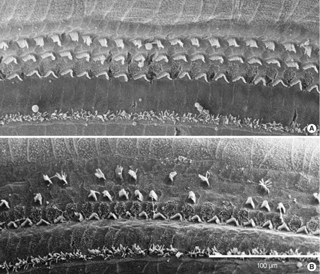
Fig. 4.

Scanning electron microscope images of organs of Corti: continuous noise exposure group (A) and repetitive noise exposure group (B). In the continuous noise exposure group, the structures of outer hair cells in the apical turn of the organ of Corti were normal (A). However, in the repetitive noise exposure group, the loss of outer hair cells and disorganization of stereocilia among the remnant outer hair cells were observed in the apical turn of the organ of Corti (B).
