
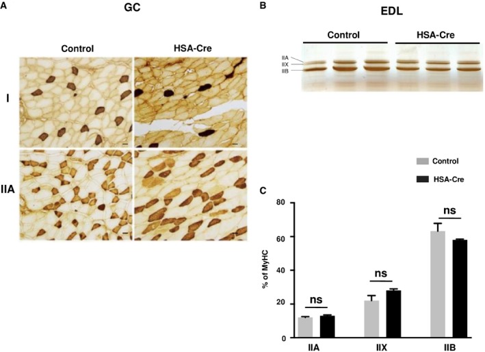
Figure EV2. Fiber type of lipin1‐deficient muscles.
-
AHistological analysis of GC by immunohistochemistry for slow (type I) and fast (type IIA). Scale bars: 40 μm.
-
B, CPanel (B) represents an electrophoretic separation of myosin heavy chain (MyHC) isoforms (IIA, IIB, IIX) in extensor digitorum longus (EDL) skeletal muscle from HSA‐Cre and control mice. Panel (C) represents the quantification of the gels in panel (B) (n = 3). Two‐tailed Student's t‐test was used for statistical analysis. Data are expressed as means ± SEM.