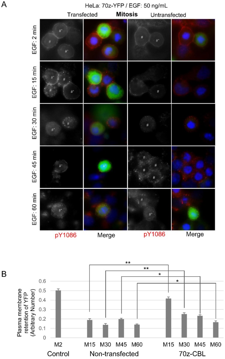
Figure 3.
Dominant-negative CBL inhibits mitotic endocytosis. Indirect immunofluorescence to observe EGFR endocytosis in HeLa cells transfected with dominant-negative CBL (70z-YFP). Following the transfection of CBL, the cells were treated with nocodazole (200 ng/mL) for 16 h. The cells were then treated with EGF (50 ng/mL) for the indicated times and were stained for pY1086 (red), and DAPI (blue). The transfected cells were green. (A) EGFR endocytosis in cells transfected with 70z-YFP. * represents interphase cells, # represents mitotic cells, and ’ represents transfected cells. (B) Quantification of EGFR retained in the plasma membrane in mitotic cells from experiments described in (A). Each data is the average of at least 10 mitotic cells. Control are mitotic cells treated with EGF (50 ng/mL) for 2 min and with fully plasma membrane-localized EGFR. The error bars are the standard errors of the mean (SEMs).

