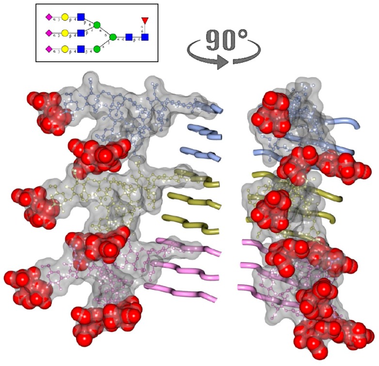
Figure 5.
Modeling of N-linked glycans in PrPSc consisting of three-rung solenoid. Two views of three-rung solenoid structures carrying tri-antennary N-glycans. Polypeptide chains are represented in the tube form, whereas N-glycans are represented in the ball-and-stick form. Each PrP molecule with corresponding N-glycan is of a different color. Sialic acid residues are colored in red. The structure of a tri-antennary N-linked glycan (shown in inset) was taken from PDB entry 3QUM, a crystal structure of human prostate specific antigen (PSA) [45]. Both calculations of electrostatic surfaces and generation of images were performed with CCP4MG software. The model based on three-rung solenoid structure is shown here for simplicity of presentation and should not be considered as preferable over the four-rung solenoid model.

