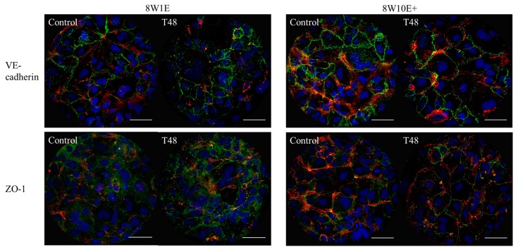
Figure 5.
Changes in the hCMVEC morphology after wounding on the 8W10E+ and 8W1E arrays using the ECIS-Zθ system. Expression of the adherens junction protein, VE-cadherin, and the tight junction regulating protein, ZO-1, under control and wounded conditions in the hCMVECs on the 8W1E and 8W10E+ arrays. Representative images of three independent experiments are shown; images are a Z-stack composition between 0.4–0.8 μm at 48 h post-wounding and the control cells. The hCMVECs are labelled for VE-cadherin using mouse monoclonal CD144 antibody, ZO-1 using mouse monoclonal ZO-1 antibody, visualized by goat α-mouse Alexa Fluor 488 (green) at 40× magnification on the LSM 710 inverted confocal microscope. Actin filaments are stained with ActinRed 555 ReadyProbes Reagent (red). Nuclei are counterstained with Hoechst (blue). The hCMVECs were seeded at a density of 60,000 cells/cm2. A wounding current of 3000 uA at 60 kHz was delivered for 30 s to selected wells on the 8W1E array, and a wounding current of 5000 uA at 60 kHz was delivered for 60 s to selected wells on the 8W10E+ array. Scale bar = 50 μm.

