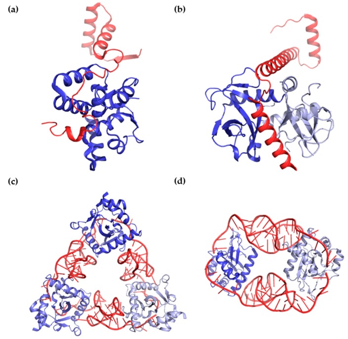
Figure 2.
Binding and interaction modes of antitoxins and toxins.; (a,b) Structural views of binding between protein antitoxins and protein toxins. (a) The antitoxin VapB26 (red) and toxin VapC26 (blue) from Mycobacterium tuberculosis interact in a 1:1 ratio (PDB code 5X3T).; (b) The antitoxin MazE4 (red) and toxin MazF4 (blue and light blue) from M. tuberculosis interact in a 1:2 ratio (PDB code 5XE3); (c,d) Structural views of binding between RNA antitoxins and protein toxins; (c) The antitoxin ToxI (red) and toxin ToxN (blue colors) from Pectobacterium atrosepticum interact in a 1:1 ratio (PDB code 2XDD); (d) The antitoxin CptI (red) and toxin CptN (blue colors) from Eubacterium rectal interact in a 1:1 ratio (PDB code 4RMO).

