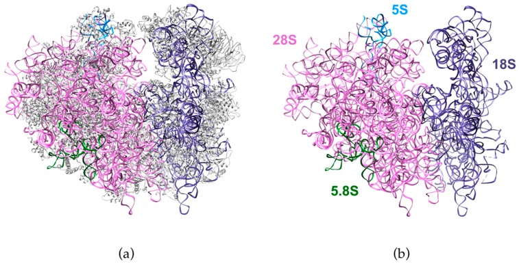
Figure 1.
Structure of the human ribosome solved by cryo-electron microscopy. The figure shows the ribosomal RNAs in the large subunit (28S: pink, 5.8S: green, 5S: blue) and in the small subunit (18S: purple). (a) Structure with the ribosomal proteins (grey). (b) Ribosomal RNAs only. Adapted from Protein Data Bank (PDB) file 4UG0 [19].

