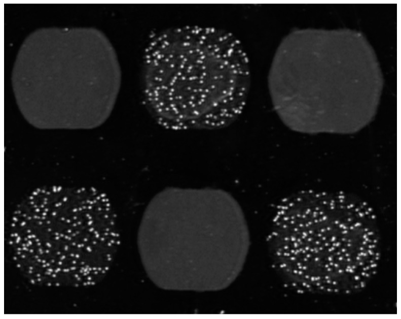
Figure 2.
Cells imaged on an SPR sensor. T-lymphocytes are visible in the SPR image on a checkerboard of anti-CD3 spots (with speckles) and BSA spots (grey areas). The image has been taken after the flow started and specific cell binding is observed on anti-CD3 spots and the not-bound cells on BSA spots were washed away. The IBIS MX96 instrument (IBIS Technologies, Enschede, the Netherlands) was used. It contains reversed optics and back and forth flow fluidics, allowing for sedimentation and controlled flow mixing of cells. Valveless injection of samples and wide 1 mm (internal diameter) tubing allows smooth aspiration of cell suspensions without clogging.

