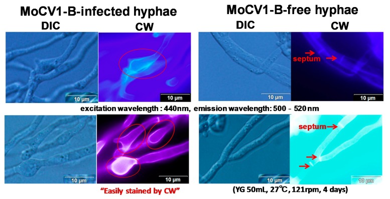
Figure 3.
Influence of MoCV1-B on cell wall formation in M. oryzae hyphae. Infected and non-infected hyphae were stained with calcofluor-white (Sigma Chemical, St. Louis, MO, USA) and examined at 1000× magnification under a light microscope (Olympus IX71, Tokyo, Japan) with differential interference contrast (DIC) optics. Calcofluor-white (CW) binds strongly to structures containing cellulose and chitin.

