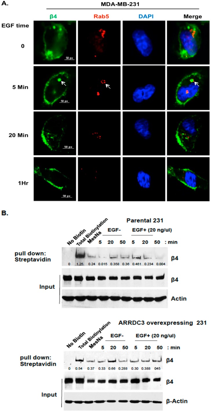
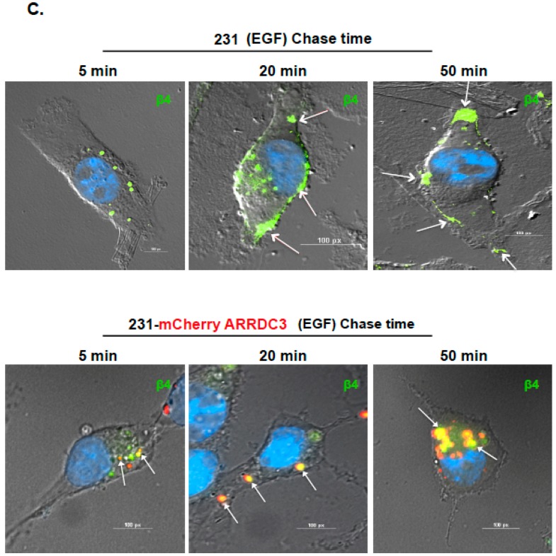
Figure 1.
ARRDC3 inhibits EGF-driven endocytic recycling of ITG β4 in triple negative breast cancer (TNBC) cells. (A) MDA-MB-231 cells were serum starved for 24 h and then stimulated with EGF (20 ng/μL) for the indicated time. Cells were stained with fluorescence tagged antibodies against ITG β4 (green) or Rab5 (Red) and DAPI (for nucleus). Arrows indicate ITG β4 inside Rab5-labled early endosomes. Scale bar: 50 μm. (B) Biotin-based recycling assay of ITG β4 was performed in MDA-MB-231 cells with or without ARRDC3 expression as described in materials and methods section. At each chase time with EGF stimulation, cells were lysed to release biotinylated proteins. Biotinylated ITG β4 were detected by immunoprecipitation (IP). Input: whole cell lysate. Representative blots of 3 independent experiments are displayed with relative input protein (Biotinylated-β4/β4 in arbitrary unit. (C) MDA-MB-231 cells were transfected with or without mCherry-ARRDC3 plasmid. Immunofluorescence-based ITG β4 recycling assay were performed as described in materials and methods. Immunofluorescence signals were captured by fluorescence microscope with DIC (differential interference contrast) optic (green; ITG β4, red; ARRDC3, yellow; co-localization). Scale bar: 100 μm. Representative images were selected from three independent experiments.


