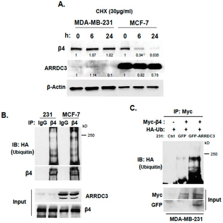
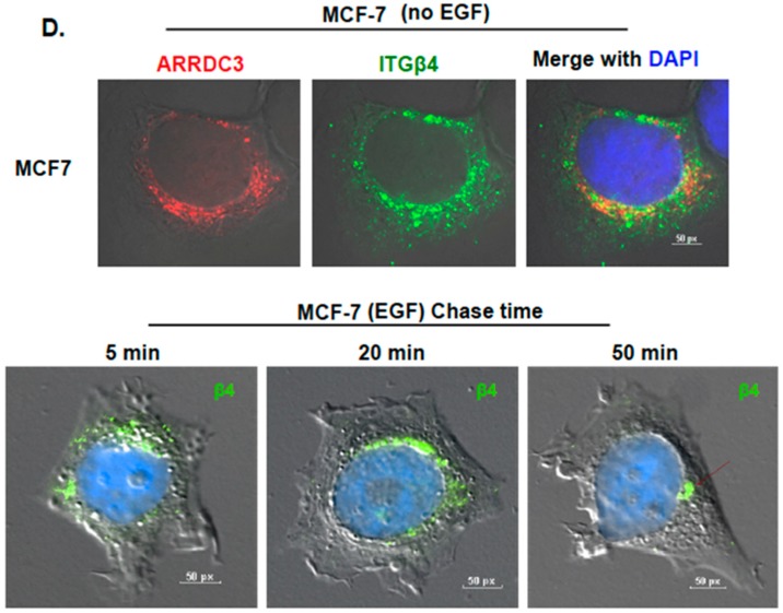
Figure 2.
Inhibition of ITG β4 recycling is linked to ARRDC3 dependent ubiquitination. (A) MDA-MB-231 and MCF-7 cells were treated with 30 μg/mL of cycloheximide (CHX) for the indicated times. The levels of ITG β4 and ARRDC3 were detected by immunoblotting (IB) analysis. β-Actin was used as a loading control. (B) Ubiquitination of ITG β4 in MDA-MB 231 and MCF-7 cells transfected with HA-Ub was detected by immunoprecipitation (IP) with ITG β4 antibody and IB with HA antibody. (C) MDA-MB-231 parental and GFP or GFP-ARRDC3 expressing MDA-MB-231 cells were co-transfected with HA-Ub and either ITG β4-Myc or Myc-empty vector. IP was performed with Myc and HA trap beads. Ubiquitination was detected by IB with HA antibody. Input; whole cell lysate. All representative images were obtained from three independent experiments. (D) MCF-7 cells were plated on cover glasses and stained with anti-ARRDC3 (red) and anti-ITG β4 (green) antibodies, followed by mounting with DAPI (blue) (upper panel). Immunofluorescence-based ITG β4 recycling assay were performed in MCF-7 cells as described in materials and methods. Immunofluorescence signals of ITG β4 (green) were captured by a fluorescence microscope with DIC optic (lower panel). Scale bar: 50 μm. Representative images were selected from three independent experiments.


