Figure 3.
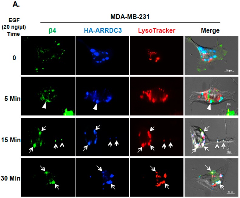
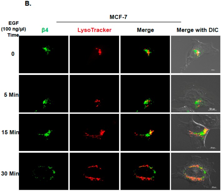
ITG β4 degradation by ARRDC3 is associated with lysosomal targeting of endosomal ITG β4. (A) MDA-MB-231 cells were transfected with HA-ARRDC3 plasmid. Next day, the cells were incubated with LysoTracker® Red DND-99 (red) before EGF treatment (20 ng/μL) for the indicated time periods. The cells were fixed and stained with fluorescence-labeled antibodies against HA (ARRDC3; blue) and ITG β4 (green). Scale bar: 50 μm. (B) MCF-7 cells were incubated with LysoTracker® Red DND-99 (red) before EGF treatment (100 ng/μL) for the indicated time periods. The cells were fixed and stained with fluorescence-labeled antibodies against ITG β4 (green). All images were captured by a fluorescence microscope with DIC optic. Scale bar: 50 μm. Representative images were selected from three independent experiments.


