Figure 6.
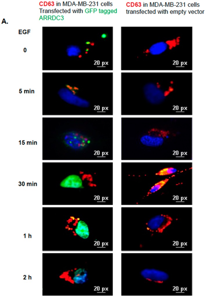
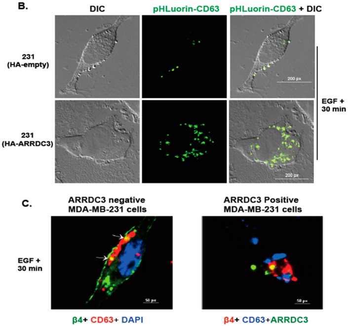
ARRDC3 prevents EGF-driven fusion of CD63 positive EVs at plasma membrane. (A) Immunofluorescence images show CD63 (red) and ARRDC3 (green) localization and DAPI nuclear staining (blue) in MDA-MB-231 cells with or without GFP-ARRDC3 for the time course with EGF treatment. Scale bar: 20 μm. (B) HA and HA-ARRDC3 expressing MDA-MB-231 cells were transfected with pHLuorin-CD63 (green) which is quenched at low pH (late endosomes) and bright at neutral pH (extracellular space or early endosome). Upon EGF treatment, CD63 location was captured by fluorescence microscope. (C) Immunofluorescence images show CD63 and ITG β4 localization in ARRDC3 positive and negative MDA-MB-231 cells at 30 min of EGF treatment (CD63; red, ITG β4; green, DAPI; blue: bottom left and CD63; blue, ITG β4; red, ARRDC3; green: bottom right). Scale bar: 50 μm. Representative images were selected from three independent experiments.


