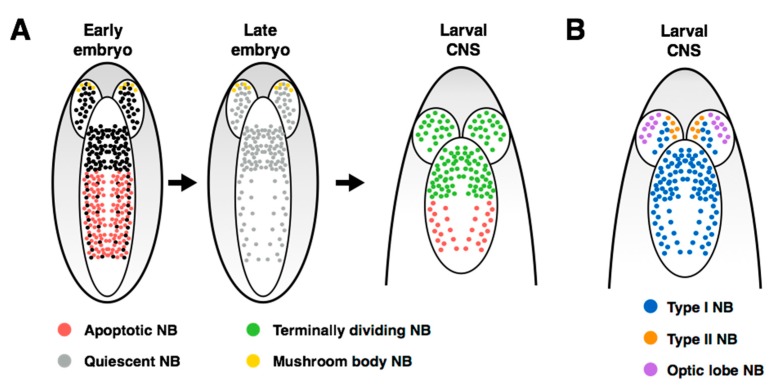
Figure 2.
Neuroblast populations within the Drosophila central nervous system. (A) Cell-fate decisions are mapped onto anatomical populations of neuroblasts during embryonic and larval development. The majority of abdominal neuroblasts undergo apoptosis during embryogenesis, leaving 3 neuroblasts per hemisegment in the larval CNS. Neuroblasts stop proliferating between embryonic and larval development, except for 4 mushroom body neuroblasts. Following reactivation, neuroblasts in the thorax and brain undergo terminal division, while the remaining abdominal neuroblasts die through apoptosis. (B) Proliferation patterns of neuroblasts throughout the larval CNS. Most neuroblasts use the Type I division pattern, except a small population of Type II neuroblasts in the brain. The neuroblasts in the inner proliferating center of the optic lobe have been observed to divide symmetrically.

