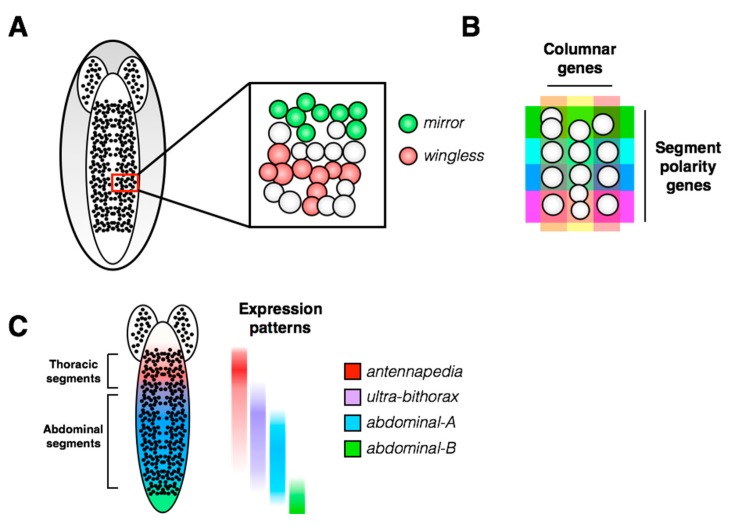
Figure 3.
Spatial patterning of the Drosophila ventral nerve cord. (A) A single hemisegment containing approximately 30 neuroblasts is shown in the inset. Gene expression is patterned within the hemisegment: two examples of spatially restricted molecular markers are shown (mirror and wingless; from the Hyper-Neuroblast Map, Doe laboratory). (B) Upon delamination from the neuroectoderm, neuroblast identity is specified within a hemisegment by a Cartesian grid generated by expression patterns of the columnar and segment polarity genes (reviewed in [16]). (C) Along the anterior-posterior axis of the embryo, hemisegment identity is also subject to spatial regulation by the Hox gene family. All of these spatial patterns are superimposed in vivo, providing each neuroblast with a unique spatial identity.

