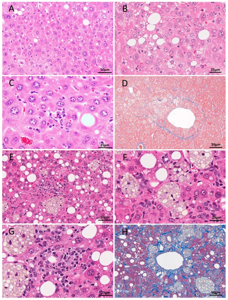
Figure 1.
H&E (A–C) and Azan (D) staining of liver sections from 52-week-old TSOD mice and H&E (E–G) and Masson trichrome (H) staining of livers in a 48-week-old mouse fed CDAHFD. In the TSOD mice livers, mild fatty change and ballooning (A,B), mild inflammation (C) and Zone 3 perisinusoidal fibrosis (D) were detected. Fatty change, lipogranuloma (E,F), severe inflammation (G) and severe fibrosis (H) were obvious in CDAHFD-fed C56Bl/6J mice. Scale bar: 25 μm (B,C,F,G), 50 μm (A,D,E,H).

