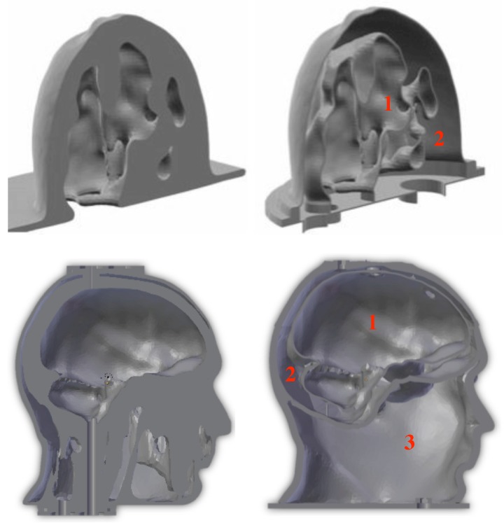
Figure 1.
Sagittal sections of the breast (up) and head (down) phantoms derived from the original STL (stereolithography) files (left) and from the modified ones (right). The red numbers indicate the various cavities that contain the different TMMs corresponding to: (1) fibroglandular or heterogeneous mix tissues, (2) fatty tissues (up-right), and (1) brain, (2) cerebrospinal fluid, (3) miscellaneous tissues (down-right).

