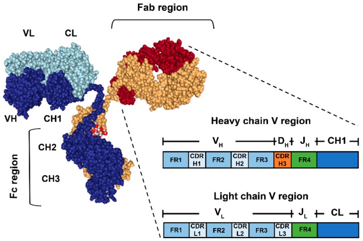
Figure 1.
Schematic representation of the basic antibody structure (Immunoglobulin G) and detailed view of a Fab. The antibody is composed of two heavy chains (dark blue and yellow) and two light chains (light blue and red). The Fab regions contain the Variable fragments of the heavy and light chains (VH and VL respectively), where the antigen-binding site is located. The zoomed in view of the Fab shows the relevant regions contained in each of the H and L chains, namely Framework 1,2, 3 and 4 (FR1,2,3,4) and Complementarity-determining Regions 1, 2 and 3 (CDRH/L1,2,3) (Not shown at scale).It is indicated as well, which of the Ig gene segments (V(D)J) codify for each of the regions after somatic recombination and rearrangement during B cell maturation. On the other hand, the Fc region contains the H chain constant segments 2 and 3 (CH2,3).

