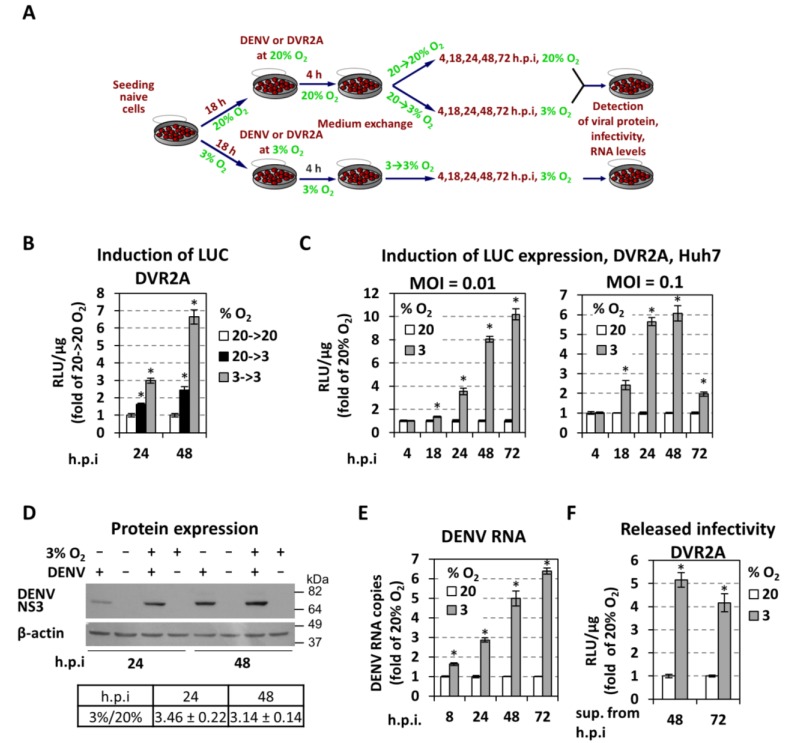
Figure 2.
Low oxygen tension enhances the production of DENV in hepatoma Huh7 cells. (A) Schematic representation of the experimental procedure. Cell culture produced DENV or DVR2a virus stocks were used for infection of naive cells that were seeded at 30% confluence (to avoid pericellular hypoxia) and preincubated for 18 h at 20% or 3% O2, respectively. After 4 h cells were washed twice with fresh culture medium, new medium was added and the incubation of cells continued as follows: cells preincubated at 20% O2 were further incubated at either 20% (referred to as 20→20% or 20%) or 3% O2 (referred to as 20→3%) whereas cells preincubated at 3% O2 were further incubated at 3% O2 (referred to as 3→3% or 3%). At the indicated time-points, cells were lysed and the expression of virus-related proteins, virus titers, and the amounts of viral RNA were determined. (B–C) Hypoxic conditions enhance DENV replication. Huh7 cells cultured under specified oxygen conditions were infected with DVR2A at MOI 0.1 (B) and MOI 0.1 or 0.01 (C), lysed at the indicated time-points and R-luc activity was measured. Values are expressed as RLU/μg of total protein amount and normalized to those obtained with 20→20% (Β) or 20% (C) O2 cells (each time-point set to one). (D) (Top) Western blot analysis of DENV NS3 protein (top) and β-actin (bottom) of DENV- and non-infected cells, incubated as specified in the top of each lane. Infection was performed with DENV at an MOI of 0.5 and cells were lysed 24 or 48 h p.i. β-actin served as a loading control. Condition of 20% O2 is indicated as “−“ and 3% O2 as “+“. Numbers on the right refer to the positions of molecular mass marker proteins. A representative experiment is shown. (Bottom) Image quantification of NS3 signals (mean values from 3 independent repetitions), normalized to β-actin and to the values obtained with cells cultured under 20% O2. (E) Viral RNA copies in cells infected with DENV at MOI = 0.01 were determined by RT-qPCR. YWHAZ mRNA levels were used for normalization. Values obtained with 20% O2 cells were set to one for each time-point. (F) Virus amounts released from Huh7 cells previously infected with DVR2A (MOI = 0.1) at the indicated oxygen conditions. Supernatants were collected at 48 and 72 h p.i. and used to infect naive Huh7 cells (infected and incubated at 20% O2), 72 h post-infection the cells were lysed and luciferase activity was measured and normalized to total protein amount. Values obtained with 20% O2 cells were set to one. In all panels, bars represent mean values from at least three independent experiments in triplicate. Error bars indicate standard deviations. * p < 0.001 vs. 20% O2 cells, for 8–72 h p.i (Student’s t-test).

