Figure 5.
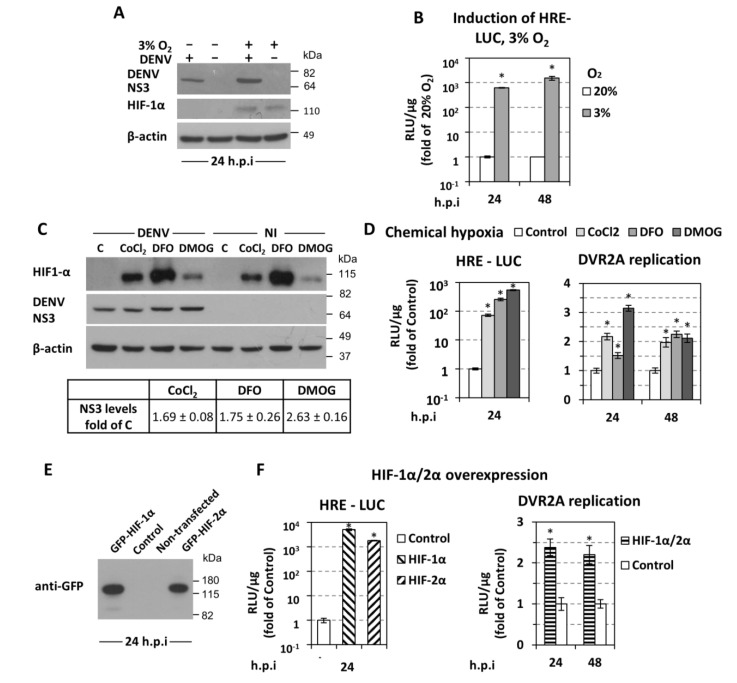
HIF upregulation enhances DENV replication. (A) Western blot analysis of DENV NS3 (top), HIF-1α (middle), and β-actin (bottom) of DENV-infected (MOI 0.5) or non-infected cells, incubated as specified on top of each lane and lysed at 24 h p.i. β-actin served as a loading control. A representative experiment of 3 independent repetitions is shown. (B) Activation of HRE (hypoxia response element) by low oxygen. Huh7 cells were transfected with the 9×HRE-Luc construct (0.4 μg/4 × 104 cells), 4 h post-transfection inoculated with DENV (MOI 0.5) for 4 h and further incubated at 20% or transferred to 3% O2 for 24 or 48 h p.i. HRE-dependent F-Luc activity was measured and expressed as RLU/μg of total protein amount. Values obtained from cells incubated at 20% O2 were set to one each time. * p < 0.001 vs. 20% O2 cells (Student’s t-test). (C,D) Chemically-induced hypoxia stabilizes HIF-1α and enhances DENV replication. (C) (Top) Western blot analysis of HIF-1α (top) DENV NS3 (middle) and β-actin (bottom) of Huh7 cells inoculated with DENV (MOI 0.5) for 4 h, and subsequently treated, for 24 h, with CoCl2 (75 μM), DFO (37.5 μM), or DMOG (62.5 μM), as specified on top of each lane. β-actin served as loading control. A representative experiment is shown. C: control non-treated cells. (Bottom) Quantification of NS3 signals from 3 independent experiments, normalized to the β-actin loading control was performed and mean values are expressed relative to that obtained from control cells. (D) Luciferase activity obtained with Huh7 cells transfected with the 9×HRE-Luc construct (left) or infected with DVR2A (MOI = 0.01, right) were treated with CoCl2 (75 μM), DFO (37.5 μM), or DMOG (62.5 μM) at 6 h post-transfection or 4 h post virus inoculation, respectively. Cells were lysed at the indicated time-points. Mean values are expressed relative to the reporter activity derived from control-non treated cells. (E,F) Overexpression of HIF-1α and HIF-2α enhances DENV replication. Huh7 cells were either co-transfected with the 9×HRE-Luc construct and a plasmid that expresses GFP-HIF-1α, GFP-HIF-2α, or empty vector (control), or first transfected with both HIF-expressing plasmids and 18 h later infected with DVR2A (MOI 0.1). (E) Western blot analysis of GFP-HIF-1α and GFP-HIF-2α using an anti-GFP antibody. (F) HRE-dependent F-Luc (left) and DVR2A-derived R-Luc (right) were measured and expressed as RLU/μg of total protein amount. Values of control cells were set to one for each time point. In all panels, bars represent mean values from at least three independent experiments in triplicate. Error bars indicate standard deviations. *p < 0.001 vs. control cells.

