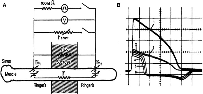
Figure 3.

Propagation of the cardiac action potential depends on current through the gap junctions between cells. (A) Schematic representation of the experimental design. (B) Electrical records in the absence of a shunt resistance (1) and presence of decreasing shunt resistance (2–6). At shunt resistance 4: the monophasic action potential changes in diphasic, meaning that threshold is reached in the right part of the preparation. Confirmation was made by the presence of a mechanical contraction (not shown). Divisions: 20 mV and 100 msec. Gap width: 400 μ (Barr et al. 1965). With permission.
