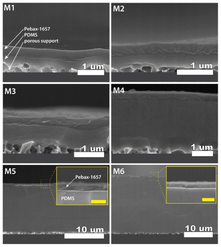
Figure 6.
Cross-section scanning electron microscopy (SEM) images (acquired on Hitachi S-5200 FESEM, with accelerating voltage 5 kV) reflecting the morphology of different fabricated TFC membranes (codes indicated in each image). All membranes containing Pebax-1657 as selective (upper) layer, PDMS as a gutter layer (middle), and porous support (bottom) as indicated for membrane M1. Insets of the cross-section show magnified views of the top Pebax-1657 layer (yellow scale bars = 1 µm). Numerical values of the thicknesses and measurement errors can be found in Table 2.

